Ocular Coherence Tomography and Alzheimer's
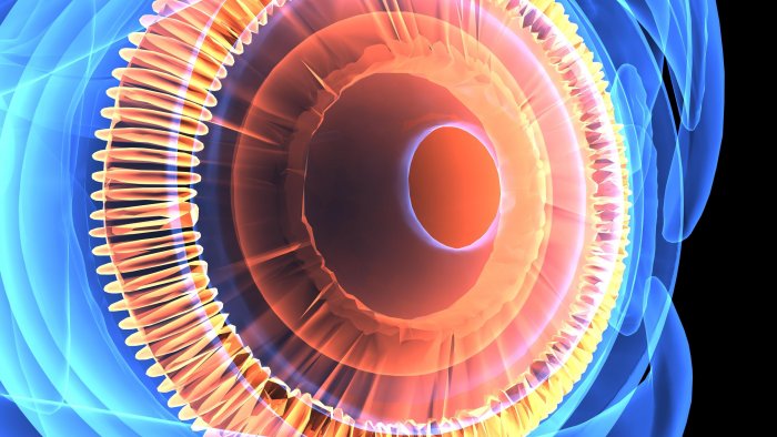
The 3D image of the retina OCT (Optical Coherence Topography) has now become a tool to study, monitor and potentially diagnose neurological disorders.
In MS, for example, the thinning of the retina can lead to optic neuritis. By defining a relationship between OCT observations and results from low-contrast vision tests, researchers have correlated a thinning retina with the vision degradation found in MS.
Though OCT is not yet incorporated into the diagnostic criteria, it has significant advantages over using magnetic resonance imaging (MRI) for diagnosing optic-nerve lesions: Its resolution is more than 1,000 times that of MRI, and OCT doesn't have the problem presented by eye movement.
OCT is also helping to uncover the early signs of Parkinson's disease and Alzheimer's disease, which could enable treatment before symptoms develop.
In November 2019, U.S, researchers combined OCT with angiography to image blood vessels. By comparing healthy retinas with those of people who had elevated levels of amyloid-β &emdash a peptide linked to Alzheimer's disease &emdash found that the foveal avascular zone, a region of the retina that lacks blood vessels, was about one-third larger, on average, in people with elevated amyloid-β
Diagnoses of Neuropsychiatric Disease
Researchers are also discovering early biomarkers of neuropsychiatric disease, which may allow clinicians to intervene before its onset or progression. For instance, there is evidence that in people with schizophrenia, the small veins, or venules, of the retina are wider and the retina is thinner.
Using electroretinography to diagnose schizophrenia
Electroretinography measures the retina's electrical response to light, and researchers at Laval University in Quebec City, Canada, are using it to identify how the response of rods and cones changes in people at risk of or diagnosed with neuropsychiatric conditions such as schizophrenia and major depressive disorder. The searchers found that the rods of young people with a high genetic risk of developing schizophrenia responded more weakly to light than those of young people without that risk.
Soon, clinicians may use the technique to distinguish between neuropsychiatric disorders. And for distinguishing between mental disorders, for example, schizophrenia and bipolar. These new tools and the ethical considerations that surround their use will continue to evolve, and perhaps become standard tools for use by the ophthalmologist.
